

3D CBCT imaging
Cone-beam 3D imaging for detailed assessment of jaw bone, impacted teeth, sinuses and implant planning, always using the smallest field of view that answers the clinical question.
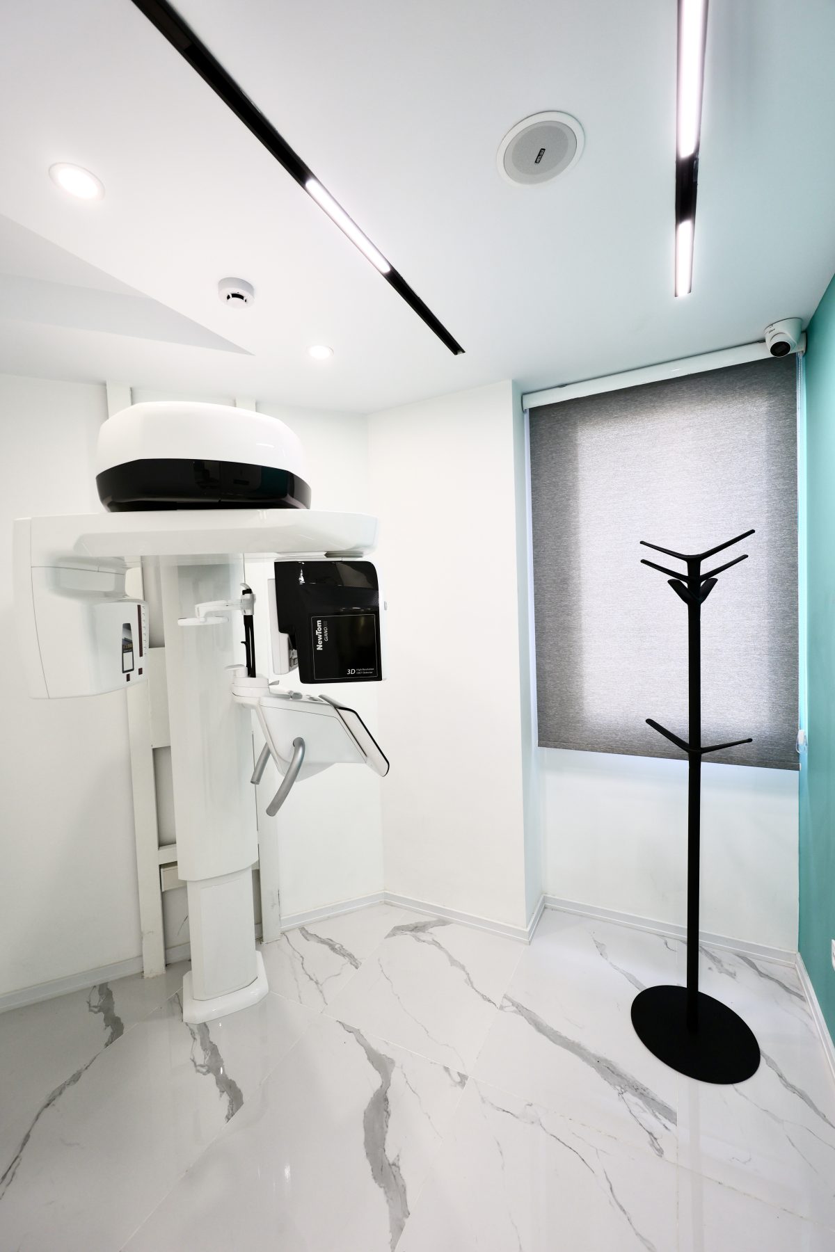
Cone Beam Computed Tomography acquires a series of images in a single rotation around the patient's head and reconstructs them into a three-dimensional model. Unlike conventional radiographs, which superimpose structures in two dimensions, CBCT lets every region be reviewed in three planes.
When is CBCT indicated?
- Implant planning and measurement of bone height and width
- Assessment of impacted teeth and their relationship to the inferior alveolar canal
- Evaluation of bone lesions, cysts and space-occupying lesions of the jaws
- Assessment of the temporomandibular joint and maxillary sinuses
- Root canal anatomy in complex endodontic cases
Choosing the field of view
The imaged volume is selected according to the diagnostic aim. We use the smallest field of view that answers the clinical question, keeping patient dose as low as reasonably achievable — the ALARA principle.
Before your visit
No special preparation is required. Please remove metal items — earrings, necklaces, hair pins, glasses and removable dentures — as they create artefacts. If you are, or may be, pregnant, please tell our staff before the examination.