


What is CBCT, and how does it differ from an ordinary dental X-ray?
CBCT builds a three-dimensional image of the jaws and teeth, allowing structures to be reviewed in three planes. Here is...
Unit 10, 2nd Floor, Bostan Building, No. 297, between Bostan 9th & 10th, Pasdaran, Tehran, Iran
Dr. Shahin Hemmati’s centre provides 3D and 2D oral and maxillofacial imaging on modern digital equipment, using low-dose protocols and accompanied by a specialist radiologist’s report.
Access patient reports, download DICOM studies and track your referrals in the dedicated colleagues’ portal.
Enter the portalBook an appointment, learn about radiation safety and find your way to our Pasdaran, Tehran location.
Book & read the guide
Radiation safety
Open the calculatorEvery study is acquired on digital equipment with a dose-reduction protocol and reported by a specialist in oral and maxillofacial radiology.
Cone-beam 3D imaging for detailed assessment of jaw bone, impacted teeth, sinuses and implant planning, always using the smallest field of view that answers the clinical question.
Read more
A single image covering both jaws, the teeth, the sinuses and the joints — the usual starting point for general dental and orthodontic assessment.
Read more
Standardised lateral and PA skull imaging for analysing skeletal relationships — the basis of orthodontic and orthognathic treatment planning.
Read more
High-detail digital intraoral imaging of individual teeth, their roots and the surrounding bone — the lowest-dose dental imaging available.
Read more
Assessment of the bony structures of the jaw joint in closed and open positions, for joint pain, joint noises and limited mouth opening.
Read more
Precise bone height and width measurement, distances to the nerve canal and sinus floor, and cross-sectional reporting ready for the referring surgeon.
Read moreEvery study is reviewed by a specialist in oral and maxillofacial radiology and issued with a written report.
The smallest field of view that answers the clinical question, in line with the ALARA principle.
Referring dentists collect reports and DICOM files from the portal — no need to visit in person.
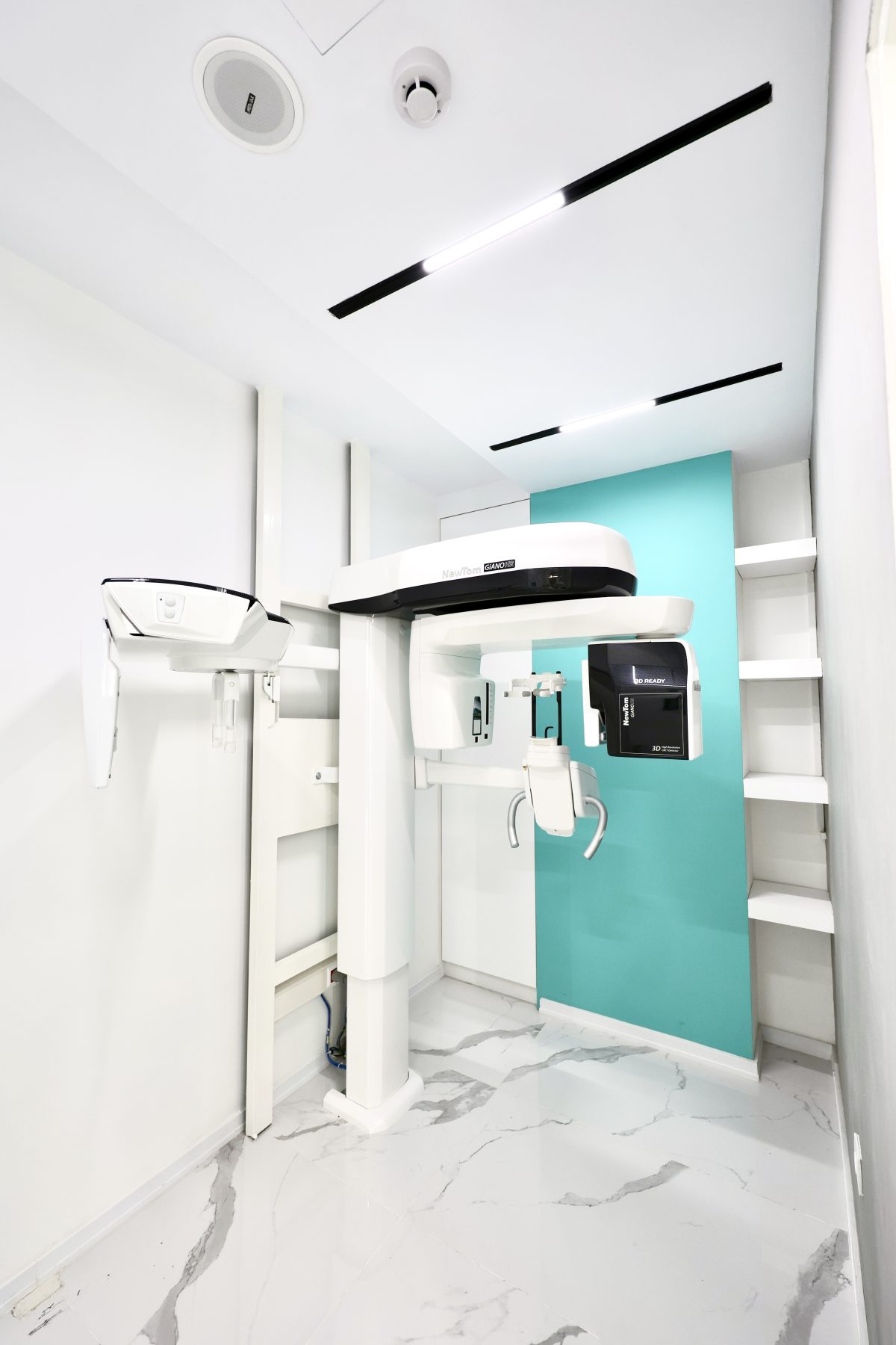
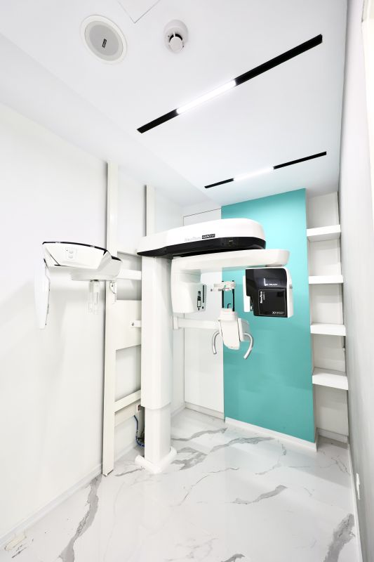
3D CBCT, digital panoramic and cephalometric imaging with high-resolution sensors.
Leave your phone number and our reception team will call you shortly to arrange a time.
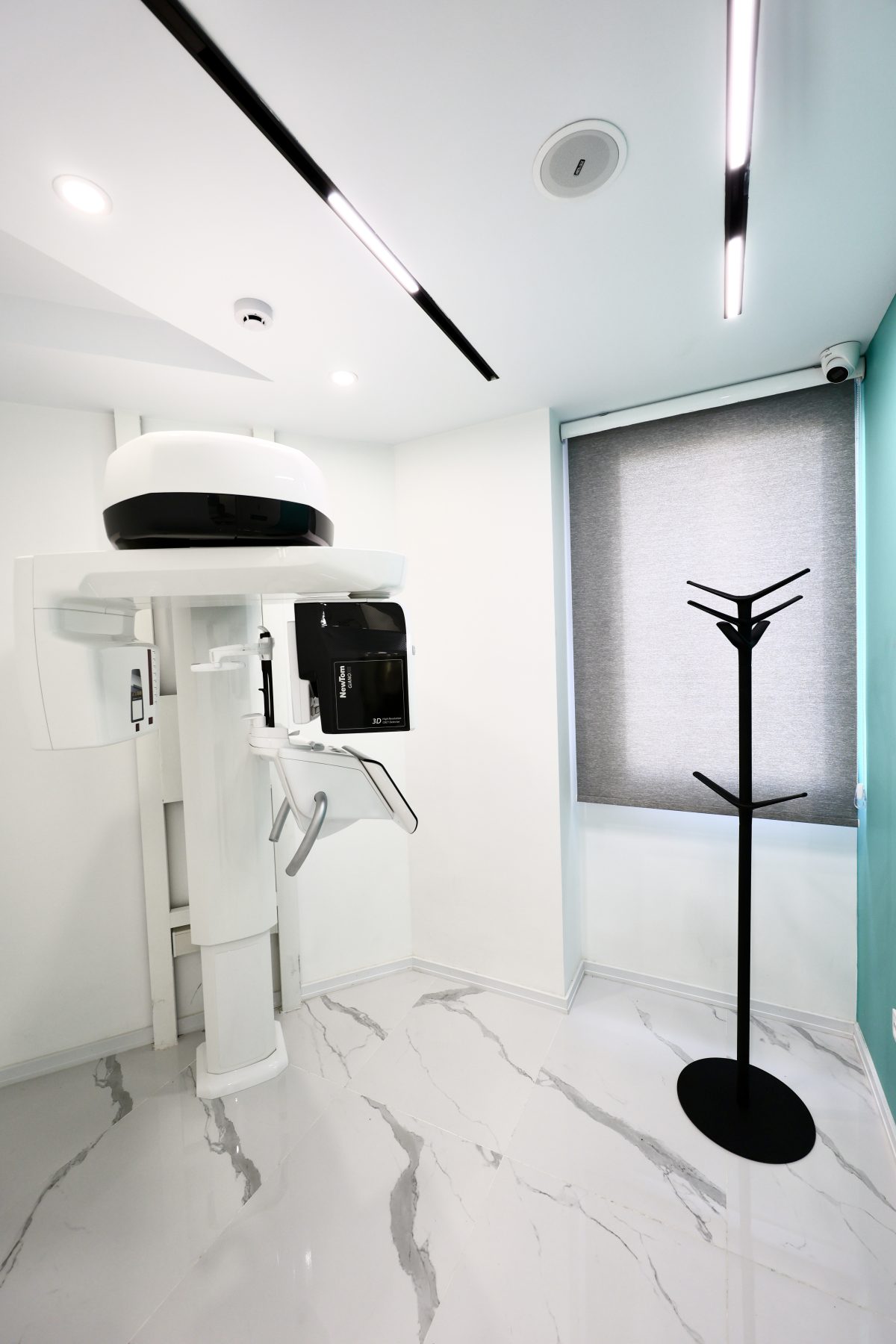
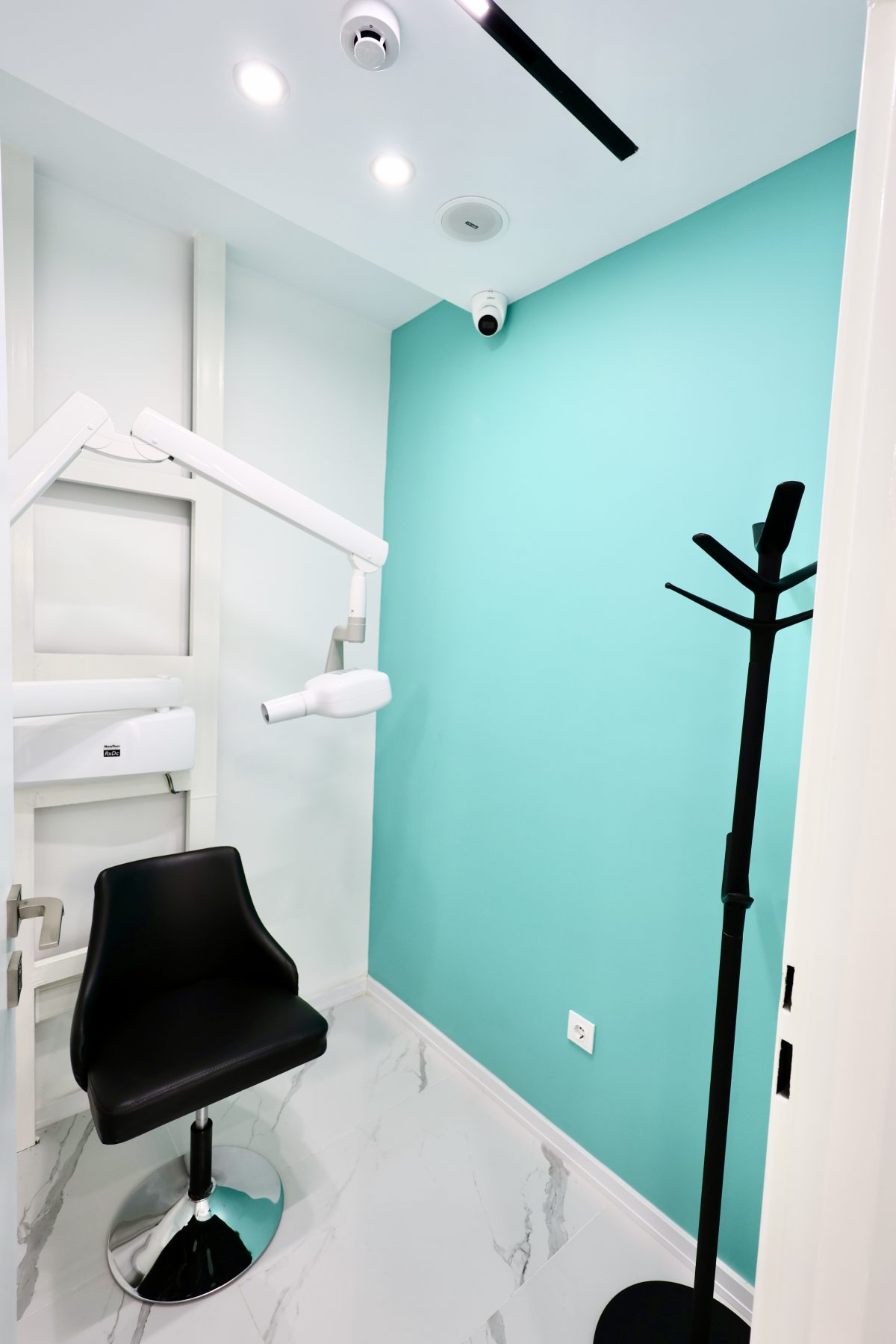
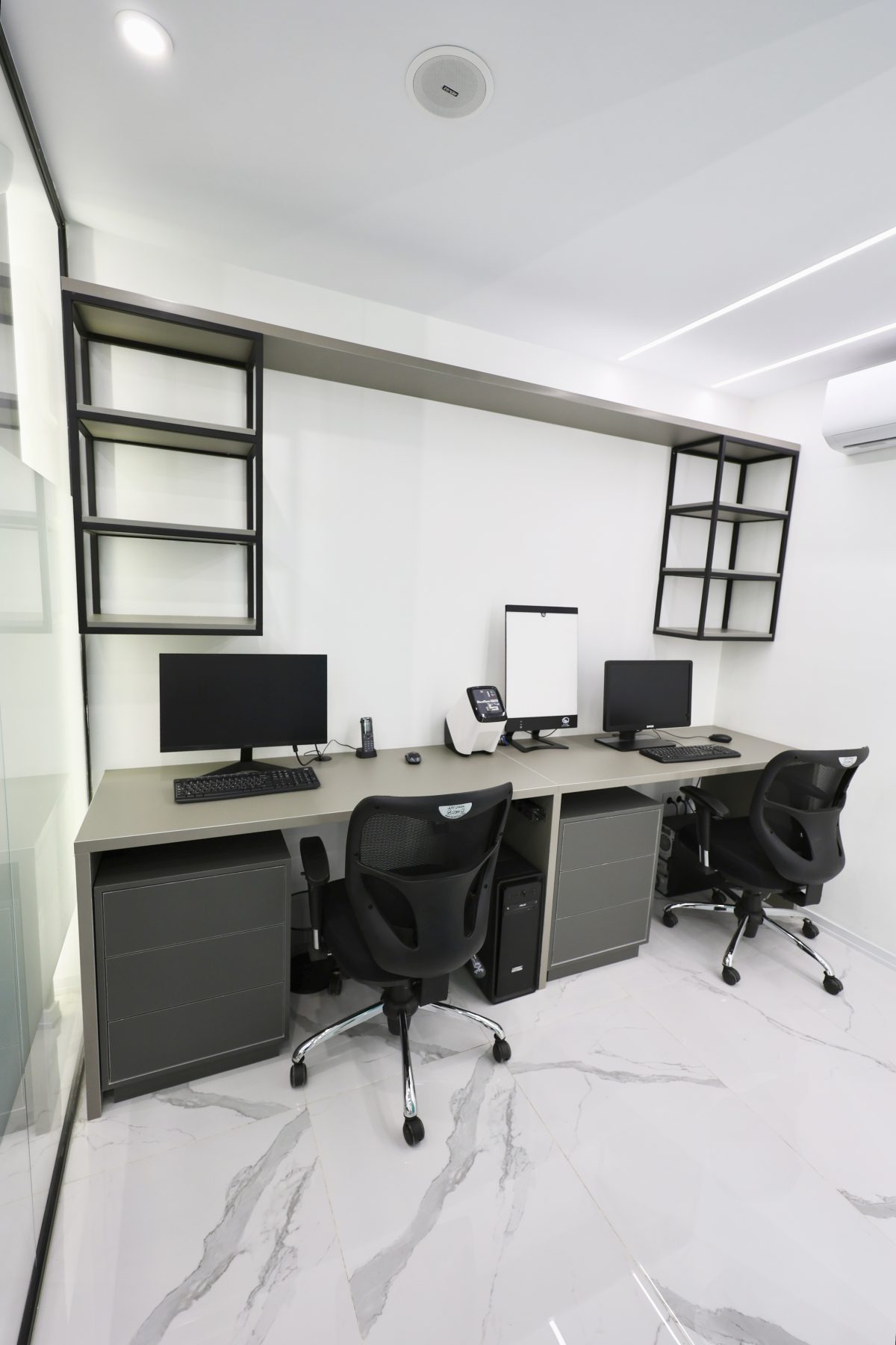
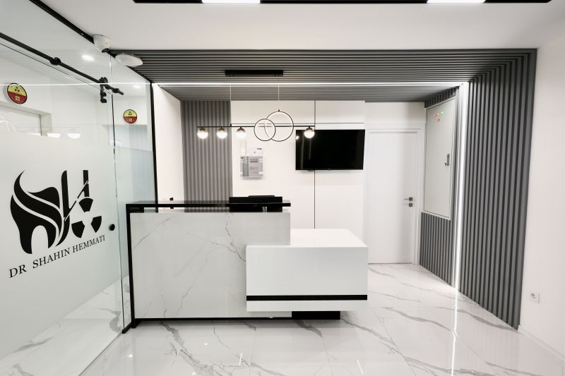
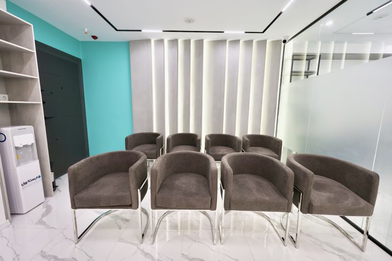
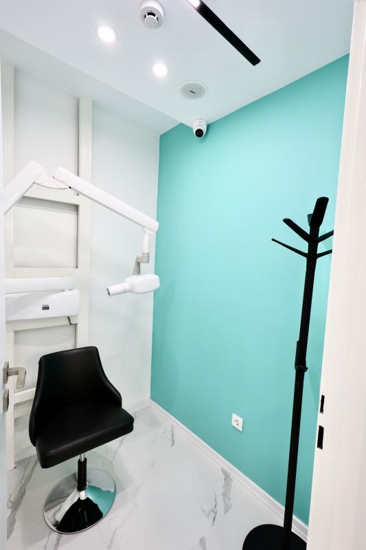
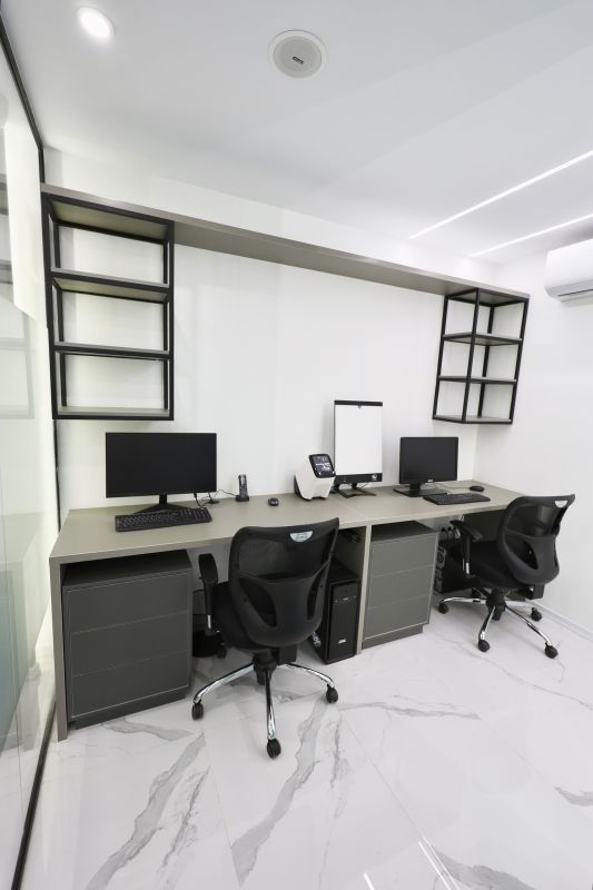
From reception to the imaging rooms, the centre is designed around radiation protection standards.
Use the interactive calculator to compare your imaging dose with natural background radiation and flight hours.
Open the calculator16 µSv
Panoramic
50 µSv
Small-FOV CBCT
6.6 µSv
Background, per day
4 µSv
Per flight hour
Answers to the questions patients and colleagues ask most about oral and maxillofacial radiography.
CBCT builds a three-dimensional image of the jaws and teeth, allowing structures to be reviewed in three planes. Here is...
Dose figures only mean something when compared with something familiar. Here we compare dental imaging doses with natura...
The quality of a radiology report depends directly on the quality of the referral. Here is what a good imaging request s...
Check our basic and supplementary insurance contracts before you visit.
18 insurance partners
InsuranceUnit 10, 2nd Floor, Bostan Building, No. 297, between Bostan 9th & 10th, Pasdaran, Tehran, Iran
Get directionsCommon questions about imaging, preparing for your visit and radiation safety.
Please bring the written request from your dentist or physician, your national ID card, and — if you are using insurance — your insurance booklet or referral letter. If you have relevant previous images, bringing them helps with comparison and a more accurate report.
No fasting or special preparation is needed. Simply remove metal items — earrings, necklaces, hair pins, glasses, hearing aids and removable dentures — before the scan, as they create artefacts on the image.
In most cases the images are available immediately after the scan, and the specialist radiologist's written report follows shortly afterwards. For urgent cases, please arrange this with reception before your visit.
DICOM is the standard medical imaging format. It holds the raw scan data and allows three-dimensional review in specialist software. Referring dentists can download it from the colleagues' portal; patients can obtain it from reception.
Doses from modern digital dental imaging are very low. A panoramic radiograph, for example, is roughly equivalent to the natural background radiation you receive over two to three days. Even so, every examination must be clinically justified, and that decision rests with your treating clinician. Use the radiation calculator on this site for a fuller comparison.
If you are, or may be, pregnant, please tell our staff and your treating clinician before the examination. During pregnancy, imaging is performed only where clinically necessary and with all protective measures in place.