

CBCT before a dental implant: why it is needed and what it shows
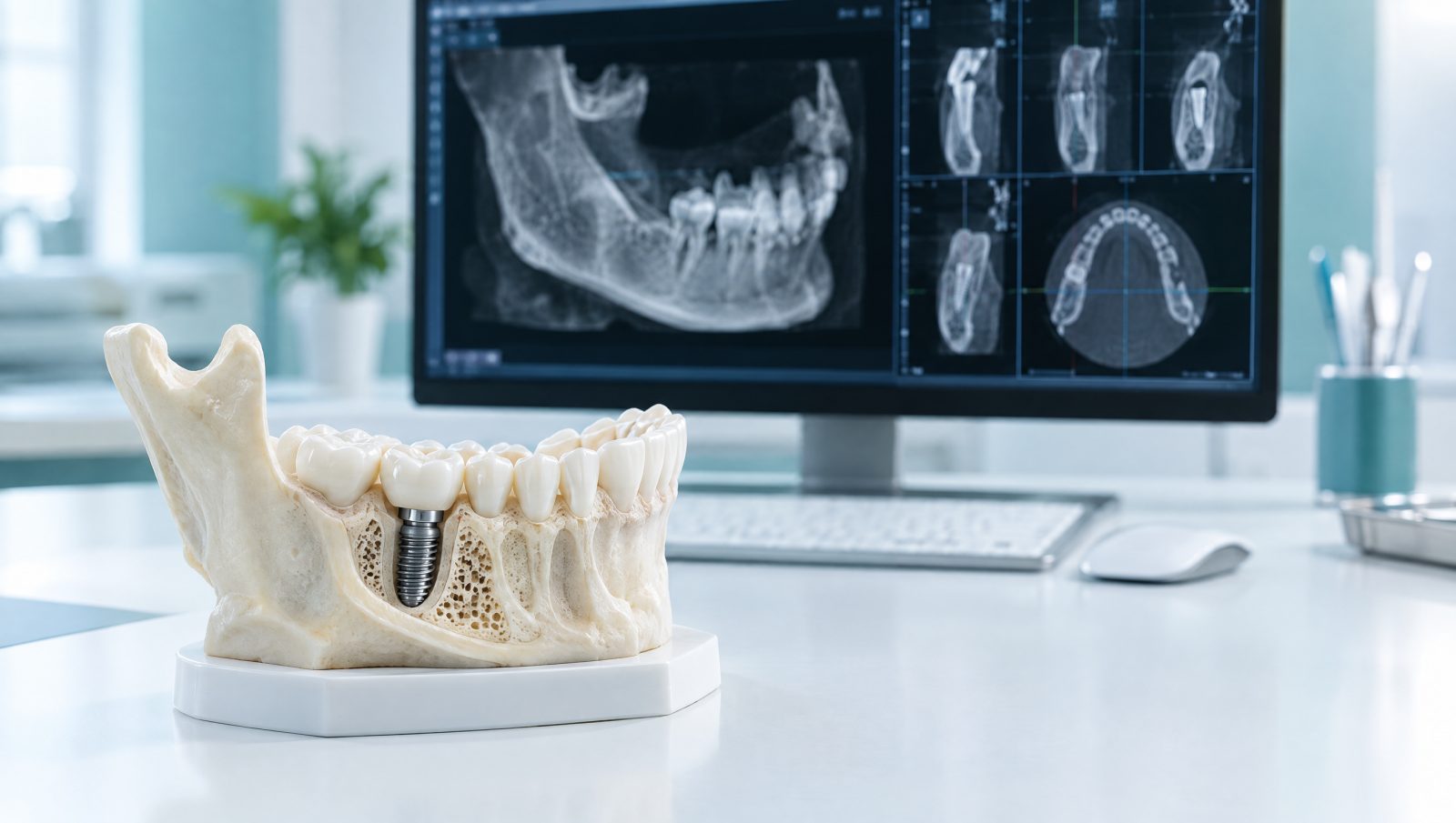
Before an implant, CBCT shows bone height and width, the course of the lower-jaw nerve and the sinus floor in three dimensions. Here is what the scan tells your clinician and how to prepare.
If you are about to have a dental implant, your dentist or surgeon will very likely request a CBCT scan first — what many patients call a "dental CT scan for implants". This three-dimensional image shows the height, width and shape of the bone at the implant site, and exactly where sensitive structures such as the nerve in the lower jaw and the maxillary sinus lie. This article explains why CBCT matters before an implant, what it shows and how to prepare.
Why a panoramic X-ray is not enough for implant planning
A panoramic X-ray is excellent for an overview, but it is two-dimensional. It shows bone height to some extent, but not the width of the bone from cheek to tongue, which is critical for choosing the implant's diameter and angle. Magnification also varies across a panoramic image, so it cannot be measured precisely. CBCT has neither limitation: it shows the bone in three planes at true scale.
What a pre-implant CBCT shows
Bone height and width
The key question before an implant is whether there is enough bone to hold it. Cross-sectional CBCT slices let the clinician measure bone height and width at every point, choose a suitable implant length and diameter, or know in advance that a bone graft will be needed.
Distance to the nerve canal in the lower jaw
In the lower jaw, the nerve that supplies sensation to the lip and chin runs through a canal in the bone and exits near the premolars through the mental foramen. Injury to this nerve can cause numbness or altered sensation of the lip and chin. CBCT shows the canal and the foramen in three dimensions so that a safe distance can be kept.
The floor of the maxillary sinus
Around the upper molars the maxillary sinus is often close to the roots, and after an extraction the bone beneath it may shrink. CBCT shows how much bone remains below the sinus and the condition of its lining, which helps decide whether a sinus lift is needed. See our maxillofacial sinus assessment service for more.
Bone shape and angle
The jawbone is not always straight and even — for example, many people have a hollow on the inner surface of the lower jaw near the molars. Seeing these contours before surgery helps avoid perforating the bone wall.
Neighbouring teeth and other findings
The position of adjacent roots, retained root fragments, cysts or bony lesions and sinus inflammation can all appear on a CBCT and change the treatment plan.
The right field of view: as much as needed, no more
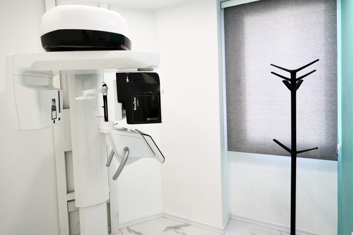
The field of view (FOV) is the size of the region imaged. One or two implants in one area need only a small field of view; multiple implants or a full-arch reconstruction need a larger one. A smaller field of view means a lower dose and usually finer detail, which is why we always choose the smallest field of view that answers the clinician's question. Referring dentists can find tips on writing a precise request in our referral guide for colleagues.
From CBCT to digital implant planning
A CBCT is more than a few printed images. The raw data are delivered in the standard DICOM format so they can be opened in implant-planning software, where the implant's position, angle and size can be set virtually before surgery. Combined with an intraoral scan, the CBCT can also be used to make a surgical guide. Learn more on our implant assessment and planning page.
Preparation and what to expect
- Your dentist's request: bring the written request, ideally naming the teeth or region of interest.
- Radiographic guide: if your dentist has made a radiographic guide or scanning stent for you, bring it.
- Metal items: remove earrings, necklaces, hair clips, glasses and removable dentures before entering the scan room.
- Staying still: the scan usually takes less than a minute, and you must stay completely still. A cotton roll or bite block is sometimes used to keep the teeth apart.
- Pregnancy: implant planning is elective and is usually postponed until after delivery. Tell us if you are, or may be, pregnant.
Radiation dose of an implant CBCT
CBCT dose depends on the field of view and the unit's settings. A small-field CBCT of one region is typically about 50 µSv — roughly a week of natural background radiation; a full-jaw CBCT is about 100 µSv and a large craniofacial CBCT about 150 µSv. These are higher than a panoramic X-ray but far lower than a conventional medical CT scan. Compare the figures with the radiation calculator.
Is CBCT needed after an implant?
Routine implant follow-up is usually done with two-dimensional images such as periapicals. A post-implant CBCT is generally requested only in specific situations — for example, unusual pain or numbness after surgery. The metal of an implant also creates streaks and shadows on CBCT that can hide nearby detail, so the decision rests with your clinician.
Why the radiologist's report matters
A CBCT produces a large amount of information, not all of it at the implant site. An oral and maxillofacial radiologist takes the measurements needed for the implant and also reviews the whole imaged volume, recording findings such as sinus changes, silent lesions or anatomical variants in a specialist report that helps the clinician plan more precisely and safely.
Frequently asked questions
Is a CBCT needed before every implant?
In most cases clinicians request one for planning, but the final decision depends on your clinical situation and rests with your treating dentist.
How is CBCT different from a regular CT scan?
CBCT uses a cone-shaped beam and a single rotation designed for the teeth and jaws, giving high-detail images of bone at a much lower dose than a conventional medical CT.
How long does it take, and when is the report ready?
The scan itself usually takes under a minute. Our reception team will tell you when your report will be ready.
Can I give the CBCT files to my own dentist?
Yes. The images are provided in DICOM format, and referring dentists can also download them from the colleagues' portal.
Do existing implants or metal crowns affect the image?
Yes — metal causes streak artefacts. Mention any metal restorations in the area so the protocol can take them into account.
To book a pre-implant CBCT, use the appointment form and our team will call you to arrange a time. You can also check our contracted insurers before your visit.